Specialty

Brain Tumors

Expansive lesions of the central nervous system treated with precision microsurgery and adjuvant therapies.

Brain Tumors
01 — WHAT IT IS

An expansive lesion inside the cranial vault.

Brain tumors result from the disordered multiplication of cells in the central nervous system, forming an expansive lesion within a closed system, the cranial vault. All age groups are affected — in children there is a predominance of the lower compartment of the skull (posterior fossa) and in adults of the upper compartment (supratentorial).

There are genetically inherited diseases, such as neurofibromatosis and tuberous sclerosis, that favor their onset. The lesions are classified by their cellular characteristics — astrocytomas, oligodendrogliomas, ependymomas, meningiomas, neurinomas — and, today, also by molecular and genetic characterization, with the goal of individualizing treatment and defining the prognosis.
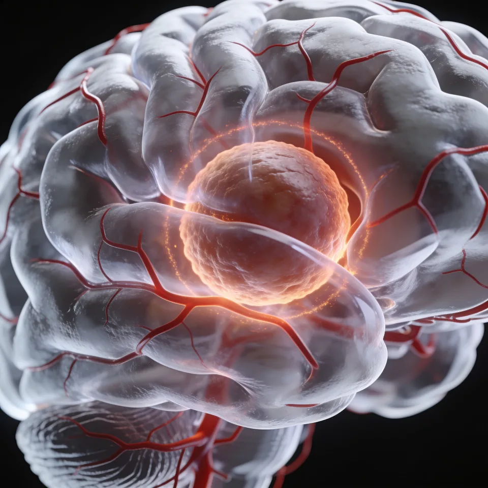
Fig. 01 — Supratentorial expansive lesion
02 — SYMPTOMS

Warning signs

The most frequent manifestations are severe headaches or a change in the pattern of the usual headache; progressive deficits and seizures may also appear:

  • Severe headache or change in pattern
  • Motor and sensory changes
  • Visual changes
  • Seizures
03 — DIAGNOSIS

Imaging diagnosis

The investigation begins with an imaging study — computed tomography and, in greater detail, MRI of the skull. After surgery, the removed tissue is analyzed and follow-up continues with neurological observation and serial MRI.

04 — TREATMENT

Management and treatment

Treatment generally begins with a surgical procedure, with the goal of collecting material for analysis and, in most cases, performing the widest possible removal with the least neurological impact:

01

Microsurgical resection

Precision microsurgical technique supported by neuronavigation, ultrasonic aspirator, and neurophysiological monitoring of sensory-motor evoked potentials to preserve eloquent areas.

02

Awake surgery and guided biopsy

Electrocorticography to identify eloquent brain areas and surgery with the patient awake; biopsies guided by stereotaxy or neuronavigation when indicated.

03

Complementary therapies

After tissue analysis, treatment may be followed by radiotherapy and chemotherapy, according to the tumor type.

“The goal is the widest possible removal, with the least neurological impact on the patient.”

Questions about your case?

Schedule an evaluation so we can discuss the diagnosis and the treatment path suited to your case.

Get in touch
Book